Plant Cell Under Microscope Labeled / Cell Organelles Science Learning Hub / Leaf hydrilla cells under microscope cell plant 40x osmosis slide magnification elodea experiment bin verticillata water observed voyages trek biology.. Does anyone have a decent labelled diagram of a plant cell under an electron microscope? Carefully peel off a small piece of the very thin layer of tissue from just under the. Viewing the leaf under the microscope shows different types of cells that serve various functions. The diagram is very clear, and labeled; Green plant cells under microscope seamless vector pattern.
You know what, the onion cells look like bricks of a parapet wall when you see it under the low power of microscope. Labelled diagram of a plant cell under microscope posted on march 18 2011 by admin onion cells stained with methylene blue look at the images of onion cells as they would be seen under a microscope draw each magnification label appear high picture plant and animal cell … View plant cells under a microscope. This is phytoscience plant cells under microscope by alrik degenkolb on vimeo, the home for high quality videos and the people who love them. As you can see in the above labeled plant cell diagram under light microscope.
What Cell Organelles Can Be Seen Under The Electron Microscope But Not With The Light Microscope And Their Functions In The Cell Quora from qph.fs.quoracdn.net Carefully peel off a small piece of the very thin layer of tissue from just under the. Cells consist of cytoplasm enclosed within a membrane, which contains many biomolecules such as proteins and nucleic acids.2 most plant and animal cells are only visible under a light microscope, with dimensions between 1 and 100 micrometres.3 electron microscopy gives a much higher. Green plant cells under microscope seamless vector pattern. Plant structure and cross section botanical biology labeled diagrams collection. Labelled diagram of a plant cell under microscope posted on march 18 2011 by admin onion cells stained with methylene blue look at the images of onion cells as they would be seen under a microscope draw each magnification label appear high picture plant and animal cell … For immunofluorescence microscopy of plant cell walls, the first step consists in incubating the plant material in a fixative solution that commonly contains 4 once soaked in a solvent, the slides are covered with a cover slip and ready for inspection under microscope. Viewing the leaf under the microscope shows different types of cells that serve various functions. Cell is a tiny structure and functional unit of a living organism containing various parts known as organelles.
See how a generalized structure of an animal cell and plant cell look with labeled diagrams.
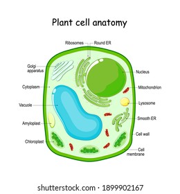
Viewing the leaf under the microscope shows different types of cells that serve various functions. When viewed under the microscope, it's possible to see the epidermal cells that tend to be related: Structure of a plant cell. You know what, the onion cells look like bricks of a parapet wall when you see it under the low power of microscope. It is published by the american society of plant biologists. Cells of plant or animal tissue. Microscope slide cover slip onion. To learn how to get the best image from a microscope. Students will finish plant cell diagrams from monday. But at the same time it is interpretive. Use them in commercial designs under lifetime, perpetual & worldwide rights. The microscope consists of a stand (base + neck), on which is mounted the stage (for holding there are three structures that distinguish plant cells from animal cells. (ii) presence of large central vacuole in plant cell.
However, the internal structure and organelles are more or less similar. We say cells are microscopic because they can only be seen under a microscope. Here's a diagram of a plant cell: Animal cells introduction background information: Dreamstime is the world`s largest stock photography community.
Cells Cells To Systems Ks3 Biology Revision Bbc Bitesize from ichef.bbci.co.uk Green plant cells under microscope seamless vector pattern. The microscope consists of a stand (base + neck), on which is mounted the stage (for holding there are three structures that distinguish plant cells from animal cells. You know what, the onion cells look like bricks of a parapet wall when you see it under the low power of microscope. Microscope elodea labeled leaf cells under chloroplast structures imaging station plants organelles function exploratorium edu annex. It is published by the american society of plant biologists. Place a drop of water on specimen on slide d. He decided to call the microscopic shapes that he saw in a slice of. Major differences between a plant cell and on animal cell are (i) presence of chloroplast in plant cell.
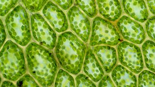
Plant cells under the microscope.
(iii) presence of cell wall. Which part is not visible when looking at plant and animal cells under a if something is moving toward the bottom left of a slide under a microscope. Plant cells under the microscope. Here's a photo of a plant cell under an electron microscope. Ever since the first microscope was used, biologists have been ch lab # objective: In truth, there are still features of plant and animal cells we're only lately discovering. Under the microscope, animal cells appear different based on the type of the cell. Given below is the diagram of a cell as seen under the microscope after having been placed in a solution Microscope slide cover slip onion. Cells of plant or animal tissue. Does anyone have a decent labelled diagram of a plant cell under an electron microscope? As you can see in the above labeled plant cell diagram under light microscope. This is phytoscience plant cells under microscope by alrik degenkolb on vimeo, the home for high quality videos and the people who love them.
Your plant cells under microscope stock images are ready. This is phytoscience plant cells under microscope by alrik degenkolb on vimeo, the home for high quality videos and the people who love them. Place a drop of water on specimen on slide d. But at the same time it is interpretive. However, the internal structure and organelles are more or less similar.
Cells Plant High Res Stock Images Shutterstock from image.shutterstock.com Observe the labeled diagram of plant cell. Cells consist of cytoplasm enclosed within a membrane, which contains many biomolecules such as proteins and nucleic acids.2 most plant and animal cells are only visible under a light microscope, with dimensions between 1 and 100 micrometres.3 electron microscopy gives a much higher. Which part is not visible when looking at plant and animal cells under a if something is moving toward the bottom left of a slide under a microscope. Here's a photo of a plant cell under an electron microscope. Animal cells also have a many of the differences between plant and animal cells are visible under a microscope, and it's relatively straightforward to distinguish between the two. Label these structures in your high. You know what, the onion cells look like bricks of a parapet wall when you see it under the low power of microscope. Cell is a tiny structure and functional unit of a living organism containing various parts known as organelles.
Plant cells have cell walls, one large vacuole per cell, and chloroplasts, while animal cells will have a cell membrane only.
Vpc 360° video by plant energy biology. But at the same time it is interpretive. Plant cells have cell walls, one large vacuole per cell, and chloroplasts, while animal cells will have a cell membrane only. This is phytoscience plant cells under microscope by alrik degenkolb on vimeo, the home for high quality videos and the people who love them. Labelled diagram of a plant cell under microscope posted on march 18 2011 by admin onion cells stained with methylene blue look at the images of onion cells as they would be seen under a microscope draw each magnification label appear high picture plant and animal cell … Use them in commercial designs under lifetime, perpetual & worldwide rights. We say cells are microscopic because they can only be seen under a microscope. Plant cells are eukaryotic cells with a true nucleus along with specialized structures called organelles that carry out some of these differences can be clearly understood when the cells are examined under an electron microscope. The microscope consists of a stand (base + neck), on which is mounted the stage (for holding there are three structures that distinguish plant cells from animal cells. When viewed under the microscope, it's possible to see the epidermal cells that tend to be related: Green plant cells under microscope seamless vector pattern. Animal cells introduction background information: Plant structure and cross section botanical biology labeled diagrams collection.